Focused-beam X-ray fluorescence and diffraction microtomographies for mineralogical and chemical characterization of unsectioned extraterrestrial samplesOPEN ACCESS
Antonio Lanzirotti, Stephen R. Sutton, Matthew Newville, Adrian Brearley, Oliver Tschauner
MAPS, Version of Record online: 17 January 2024
LINK (OPEN ACCESS)
PDF (OPEN ACCESS)
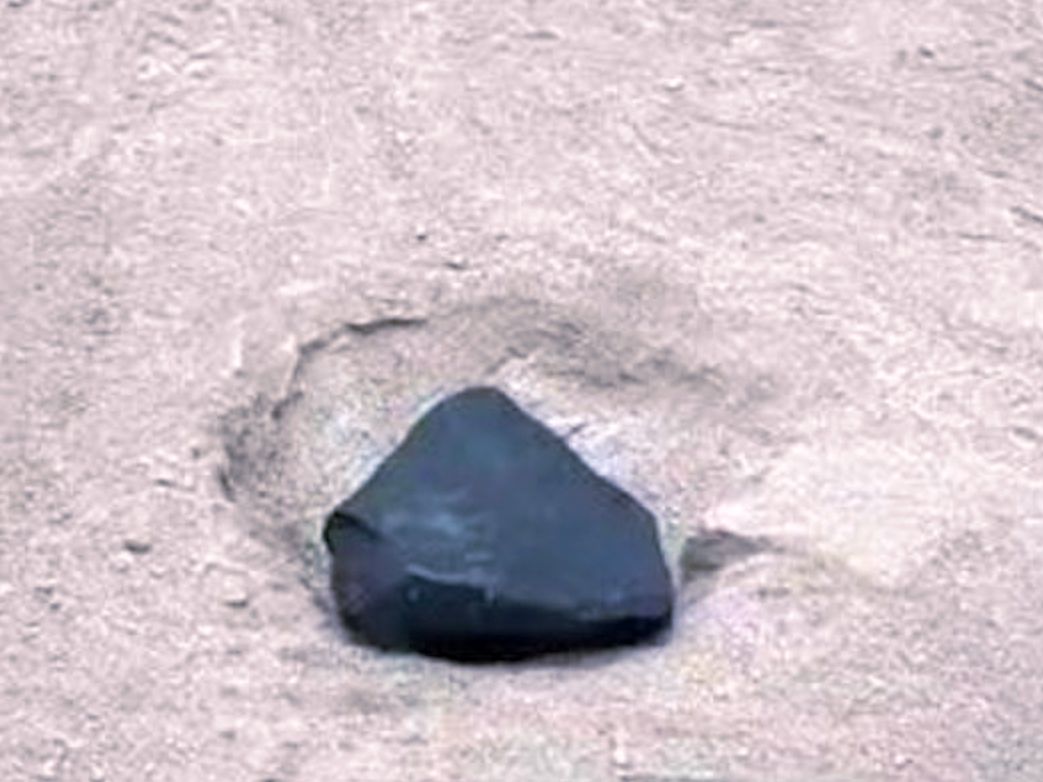
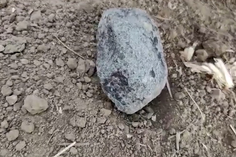
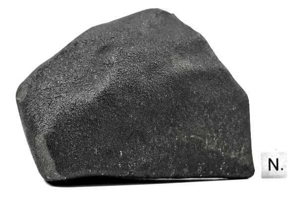
“This study describes the application of new synchrotron X-ray fluorescence (XRF) and diffraction (XRD) microtomographies for the 3-D visualization of chemical and mineralogical variations in unsectioned extraterrestrial samples. These improved methods have been applied to three compositionally diverse chondritic meteorite samples that were between 300 and 400 μm in diameter, including samples prepared from fragments of the CR2 chondrite LaPaz Icefield (LAP) 02342, H5 chondrite MacAlpine Hills (MAC) 88203, and the CM2 chondrite Murchison. The synchrotron-based XRF and XRD tomographies used are focused-beam techniques that measure the intensities of fluorescent and diffracted X-rays in a sample simultaneously during irradiation by a high-energy microfocused incident X-ray beam. Measured sinograms of the emitted and diffracted intensities were then tomographically reconstructed to generate 2-D slices of XRF and XRD intensity through the sample, with reconstructed pixel resolution of 1–2 μm, defined by the resolution of the focused incident X-ray beam. For sample LAP 02342, primary mineral phases that were visualized in reconstructed slices using these techniques included isolated grains of α-Fe, orthopyroxene, and olivine. For our sample of MAC 88203, XRF/XRD tomography allowed visualization of forsteritic olivine as a primary mineral phase, a vitrified fusion crust at the sample surface, identification of localized Cr-rich spinels at spatial resolutions of several micrometers, and imaging of a plagioclase-rich glassy matrix. In the sample of Murchison, major identifiable phases include clinoenstatite- and olivine-rich chondrules, variable serpentine matrix minerals and small Cr-rich spinels. Most notable in the tomographic analysis of Murchison is the ability to quantitatively distinguish and visualize the complex mixture of serpentine-group minerals and associated tochilinite–cronstedtite intergrowths. These methods provide new opportunities for spatially resolved characterization of sample texture, mineralogy, crystal structure, and chemical state in unsectioned samples. This provides researchers an ability to characterize such samples internally with minimal disruption of sample micro-structures and chemistry, possibly without the need for sample extraction from some types of sampling and capture media.”

