Effect of polychromatic X‐ray microtomography imaging on the amino acid content of the Murchison CM chondrite
Jon M. Friedrich, Hannah L. McLain, Jason P. Dworkin, Daniel P. Glavin, W. Henry Towbin, Morgan Hill, Denton S. Ebel
Meteoritics & Planetary Science
First published: 23 August 2018
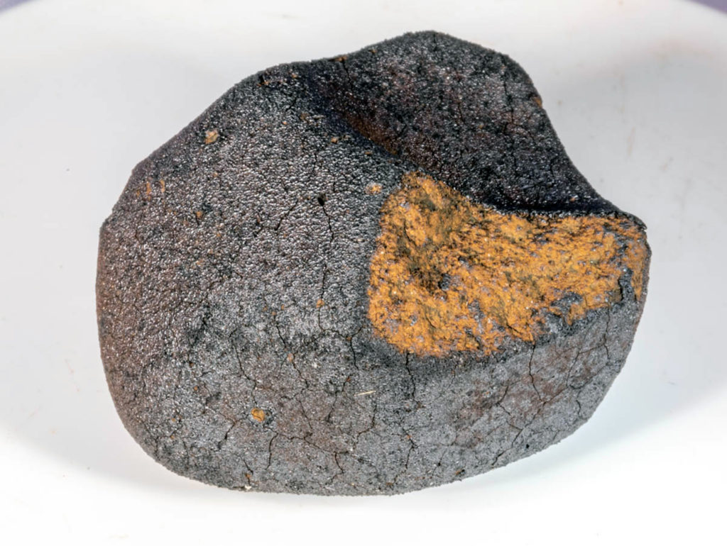
“X‐ray microcomputed tomography (μCT) is a useful means of characterizing cosmochemical samples such as meteorites or robotically returned samples. However, there are occasional concerns that the use of μCT may be detrimental to the organic components of a chondrite. Small organic compounds such as amino acids comprise up to ~10% of the total solvent extractable carbon in CM carbonaceous chondrites. We irradiated three samples of the Murchison CM carbonaceous chondrite under conditions akin to and harsher than those typically used during typical benchtop X‐ray μCT imaging experiments to determine if detectable changes in the amino acid abundance and distribution relative to a nonexposed Murchison control sample occurred. After subjecting three meteorite samples to ionizing radiation dosages between ~300 Gray (Gy) and 3 kGy with bremstrahlung X‐rays, we analyzed the amino acid content of each sample. Within sampling and analytical errors, we cannot discern differences in the amino acid abundances and amino acid enantiomeric ratios when comparing the control samples (nonexposed Murchison) and the irradiated samples. We conclude that a polychromatic X‐ray μCT experiment does not alter the abundances of amino acids to a degree greater than how well those abundances are measured with our techniques and therefore any damage to amino acids is minimal. “

